Tenure-Track Assistant Professor
University of Florida J. Crayton Pruitt Family Department of Biomedical Engineering

A health data scientist, Ruogu Fang is the Assistant Professor at University of Florida’s J. Crayton Pruitt Family Department of Biomedical Engineering in Herbert Wertheim College of Engineering. Her research spans data, brain and health. She focuses on questions such as: How to evaluate brain health, via mining the big medical data? She also explores how to make medical imaging higher quality and lower risk for the broad population. Fang’s current research is rooted in the big medical data and brain dynamics understanding. She is the recipient of the single-PI NSF CRII Award, Best Paper Award from IEEE International Conference on Image Processing, Ralph Lowe Junior Faculty Enhancement Award from ORAU, and Robin Sidhu Memorial Young Scientist Award from Society of Brain Mapping and Therapeutics, among the others.
Fang teaches courses such as “Machine Learning”, “Data Mining” and "Computer Applications to Biomedical Engineering". She also runs SMILE lab, standing for Smart Medical Informatics Learning and Evaluation, which also reflects for her expectation for every member in the lab to smile while exploring the human brain with big data.
University of Florida J. Crayton Pruitt Family Department of Biomedical Engineering
Florida International University School of Computing and Information Sciences
Ph.D. in Electrical and Computer Engineering
Cornell University, Ithaca, NY.
B.E. in Information Engineering
Zhejiang University, Hangzhou, China


Brain dynamics, which reflects the healthy or pathological states of the brain with quantifiable, reproducible, and indicative dynamics values, remains the least understood and studied area of brain science despite its intrinsic and critical importance to the brain. Unlike other brain information such as the structural and sequential dimensions that have all been extensively studied with models and methods successfully developed, the 5th dimension, dynamics, has only very recently started receiving systematic analysis from the research community. The state-of-the-art models suffer from several fundamental limitations that have critically inhibited the accuracy and reliability of the dynamic parameters’ computation. First, dynamic parameters are derived from each voxel of the brain spatially independently, and thus miss the fundamental spatial information since the brain is ?connected?. Second, current models rely solely on single-patient data to estimate the dynamic parameters without exploiting the big medical data consisting of billions of patients with similar diseases.
This project aims to develop a framework for data-driven brain dynamics characterization, modeling and evaluation that includes the new concept of a 5th dimension – brain dynamics – to complement the structural 4-D brain for a complete picture. The project studies how dynamic computing of the brain as a distinct problem from the image reconstruction and de-noising of convention models, and analyzes the impact of different models for the dynamics analysis. A data-driven, scalable framework will be developed to depict the functionality and dynamics of the brain. This framework enables full utilization of 4-D brain spatio-temporal data and big medical data, resulting in accurate estimations of the dynamics of the brain that are not reflected in the voxel-independent models and the single patient models. The model and framework will be evaluated on both simulated and real dual-dose computed tomography perfusion image data and then compared with the state-of-the-art methods for brain dynamics computation by leveraging collaborations with Florida International University Herbert Wertheim College of Medicine, NewYork-Presbyterian Hospital / Weill Cornell Medical College (WCMC) and Northwell School of Medicine at Hofstra University. The proposed research will significantly advance the state-of-the-art in quantifying and analyzing brain structure and dynamics, and the interplay between the two for brain disease diagnosis, including both the acute and chronic diseases. This unified approach brings together fields of Computer Science, Bioengineering, Cognitive Neuroscience and Neuroradiology to create a framework for precisely measuring and analyzing the 5th dimension – brain dynamics – integrated with the 4-D brain with three dimensions from spatial data and one dimension from temporal data. Results from the project will be incorporated into graduate-level multi-disciplinary courses in machine learning, computational neuroscience and medical image analysis. This project will open up several new research directions in the domain of brain analysis, and will educate and nurture young researchers, advance the involvement of underrepresented minorities in computer science research, and equip them with new insights, models and tools for developing future research in brain dynamics in a minority serving university.


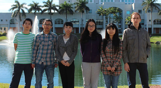
The fiber of this lab’s existence rests on both undergraduate and graduate students. We are always interested in having intelligent, motivated, and responsible students join the lab.
Smart learning and evaluation has revolutionized traditional medical data computing paradigm by converging machine learning, computer vision, big-data strategy and data mining into the conventional medical and clinical world. It has opened up tremendous opportunities in patient-centric medical imaging, robust neural computing, personal health tracking, and large-scale health informatics, etc. The medical informatics ranges over image, video, audio, text and various modalities such as CT, MRI, PET/CT, EEG and so on.
Smart Medical Informatics Learning and Evaluation (SMILE) Group focuses on scientific approaches to bridge data and medicine. Our effort spans from safety-aware medical imaging, robust hemodynamics estimation, to innovation of the computing and analysis algorithms. In our medical informatics learning and evaluation research, we explore and leverage the large amount of medical data in various forms and modalities, to generate scientific enlightening or clinically useful information. With these words, we aim to push the fundamental limits of the medical analysis and to revolutionize the functional imaging and data interpretation.
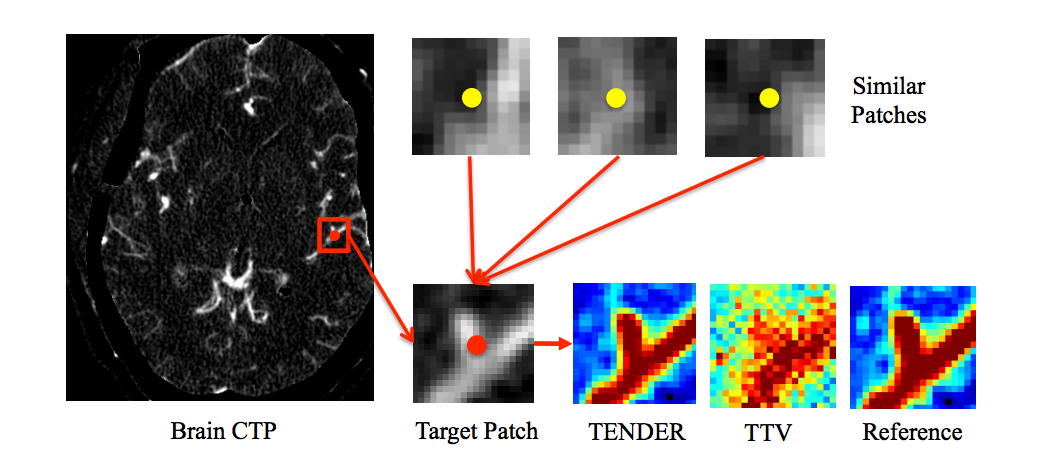
Stroke is the leading cause of long-term disability and the second leading cause of mortality in the world, and exerts an enormous burden on the public health. CT remains one of the most widely used imaging modality for stroke diagnosis. However when coupled with CT perfusion, the excessive radiation exposure in repetitive imaging to assess treatment response and prognosis has raised significant public concerns regarding its potential hazards to both short- and longterm health outcomes. Tensor total variation has been proposed to reduce the necessary radiation dose in CT perfusion without comprising the image quality by fusing the information of the local anatomical structure with the temporal blood flow model. However the local search in the framework fails to leverage the non-local information in the spatio-temporal data. In this paper, we propose TENDER, an efficient framework of non-local tensor deconvolution to maintain the accuracy of the hemodynamic parameters and the diagnostic reliability in low radiation dose CT perfusion. The tensor total variation is extended using non-local spatio-temporal cubics for regularization to integrate contextual and non-local information. We also propose an efficient framework consisting of fast nearest neighbor search, accelerated optimization and parallel computing to improve the efficiency and scalability of the non-local spatio-temporal algorithm. Evaluations on clinical data of subjects with cerebrovascular disease and normal subjects demonstrate the advantage of non-local tensor deconvolution for reducing radiation dose in CT perfusion.
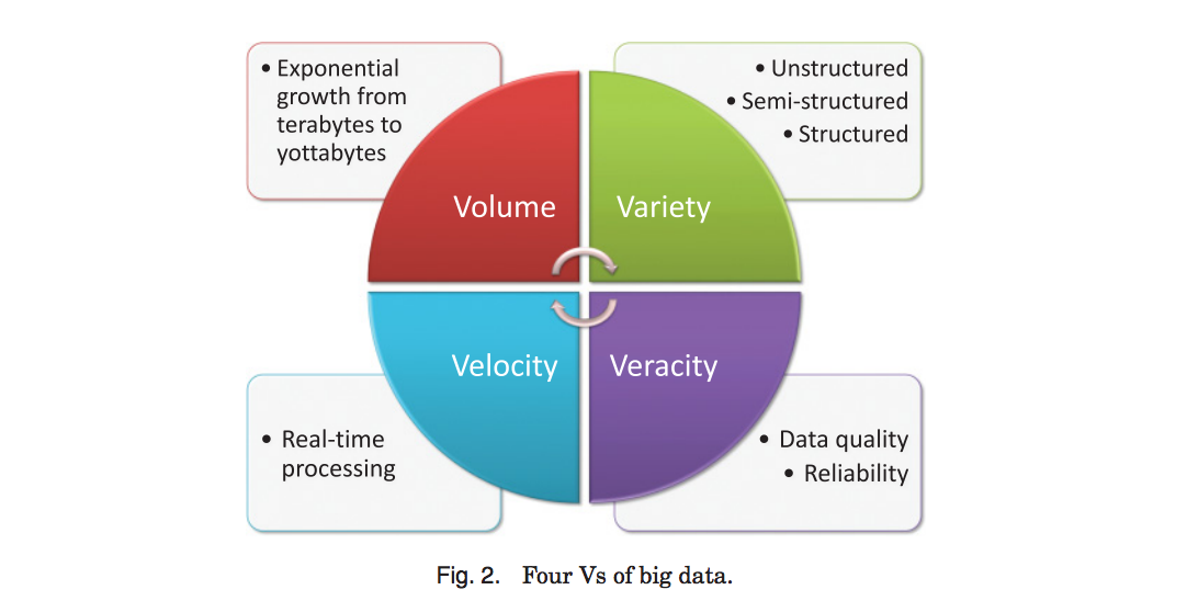
The explosive growth and widespread accessibility of digital health data have led to a surge of research activity in the healthcare and data sciences fields. The conventional approaches for health data management have achieved limited success as they are incapable of handling the huge amount of complex data with high volume, high velocity, and high variety. This article presents a comprehensive overview of the existing challenges, techniques, and future directions for computational health informatics in the big data age, with a structured analysis of the historical and state-of-the-art methods. We have summarized the challenges into four Vs (i.e., volume, velocity, variety, and veracity) and proposed a systematic data-processing pipeline for generic big data in health informatics, covering data capturing, storing, sharing, analyzing, searching, and decision support. Specifically, numerous techniques and algorithms in machine learning are categorized and compared. On the basis of this material, we identify and discuss the essential prospects lying ahead for computational health informatics in this big data age.
Near-Infrared (NIR) optical imaging can reveal tissue oxygenation of the wound, complementing the visual inspection of the surface granulation. Herein, graph cuts algorithm is applied to segment NIR images of the wound from its peripheries.

The Behavioral Risk Factor Surveillance System (BRFSS) is the nation's premier system of health-related telephone surveys that collect state data about U.S. residents regarding their health-related risk behaviors, chronic health conditions, and use of preventive services. Established in 1984 with 15 states, BRFSS now collects data in all 50 states as well as the District of Columbia and three U.S. territories. BRFSS completes more than 400,000 adult interviews each year, making it the largest continuously conducted health survey system in the world.
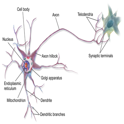
We are going to analysize different neuron from many species by data mining and deep learning algorithm seperately.
Timely detection and treatment of microaneurysms (MA) is a critical step to prevent the development of vision-threatening eye dis- eases such as diabetic retinopathy. However, detecting MAs in fundus images is a highly challenging task due to the large variation of imag- ing conditions. In this paper, we focus on developing an interleaved deep mining technique to cope intelligently with the unbalanced MA detection problem. Specifically, we present a clinical report guided multi-sieving convolutional neural network (MS-CNN) which leverages a small amount of supervised information in clinical reports to identify the potential MA regions via a text-to-image mapping in the feature space. These potential MA regions are then interleaved with the fundus image information for multi-sieving deep mining in a highly unbalanced classification problem. Critically, the clinical reports are employed to bridge the semantic gap between low-level image features and high-level diagnostic information. Extensive evaluations show our framework achieves 99.7% precision and 87.8% recall, comparing favorably with the state-of-the-art algorithms. Integration of expert domain knowledge and image information demon- strates the feasibility to reduce the training difficulty of the classifiers under extremely unbalanced data distribution.
Computed tomography perfusion (CTP) is one of the most widely used imaging modality for cerebrovascular disease diagnosis and treatment, especially in emergency situations. While cerebral CTP is ca- pable of quantifying the blood flow dynamics by continuous scanning at a focused region of the brain, the associated excessive radiation increases the patients’ risk levels of developing cancer. To reduce the necessary radiation dose in CTP, decreasing the temporal sampling frequency is one promising direction. In this paper, we propose STAR, an end-to- end Spatio-Temporal Architecture for super-Resolution to significantly reduce the necessary scanning time and therefore radiation exposure. The inputs into STAR are multi-directional 2D low-resolution spatio- temporal patches at different cross sections over space and time. Via training multiple direction networks followed by a conjoint reconstruc- tion network, our approach can produce high-resolution spatio-temporal volumes. The experiment results demonstrate the capability of STAR to maintain the image quality and accuracy of cerebral hemodynamic parameters at only one-third of the original scanning time.
Cardiovascular disease is one of the leading causes of death in the United States. It is critical to identify the risk factors associated with cardiovascular diseases and to alert individuals before they experience a heart attack. In this paper we propose RFMiner, a risk factor discovery and mining framework for identifying significant risk factors using integrated measures. We provide the blueprints for accurately predicting the possibility of heart attacks in the near future while identifying notable risk factors - especially the factors which are not well recognized.
Morphological retrieval is an effective approach to explore large-scale neuronal databases, as the morphology is correlated with neuronal types, regions, functions, etc. In this paper, we focus on the neuron identification and analysis via morphological retrieval. In our proposed framework, multiple features are extracted to represent 3D neuron data. Because each feature reflects different levels of similarity between neurons, we group features into different hierarchies to compute the similarity matrix. Then, compact binary codes are generated from hierarchical features for efficient similarity search. Since neuronal cells usually have tree-topology structure, it is hard to distinguish different types of neurons simply via traditional binary coding or hashing methods based on Euclidean distance metric and/or linear hyperplanes. Therefore, we employ an asymmetric binary coding strategy based on the maximum inner product search (MIPS), which not only makes it easier to learn the binary coding functions, but also preserves the non-linear characteristics of the neuron morphological data. We evaluate the proposed method on more than 17,000 neurons, by validating the retrieved neurons with associated cell types and brain regions. Experimental results show the superiority of our approach in neuron morphological retrieval compared with other state-of-the-art methods. Moreover, we demonstrate its potential use cases in the identification and analysis of neuron characteristics from large neuron databases.
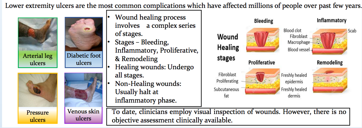
Clinicians employ visual inspection of the wound and reduction in its size over time to monitor its healing process. Although these are standard clinical assessments, there is a need to develop a physiological approach that can map sub-surface tissue oxygenation at and around the wound region. Recently, a non-contact, portable, hand-held near infrared optical scanner (NIROS) has been developed to functionally image wound sites and differentiate healing from non-healing in lower extremity ulcers. Past studies using NIROS focused on differentiating healing from non-healing wounds based on NIR optical contrast between the wound and healthy surrounding tissue. However, these studies did not showcase the physiological changes in tissue oxygenation. Herein, NIROS has been modified to perform multi wavelength imaging in order to obtain the oxy and deoxy- hemoglobin maps of the wound and its surroundings. Clinical studies are currently performed at two clinical sites in Miami on lower extremity ulcers (2, diabetic foot ulcers (DFUs) and 4 venous leg ulcers (VLUs to date). Preliminary results have shown changes in oxy- and deoxy-hemoglobin maps of the wound and background across weeks of the treatment process. Image segmentation studies quantified regions of varied tissue oxygenation around and beneath the wound to potentially determine sub-surface healing regions. Ongoing efforts involve systematic 8-week imaging studies to obtain physiological indicators of healing from hemodynamic studies of DFUs and VLUs.
Segmentation of abdominal adipose tissues (AAT) into subcutaneous adipose tissues (SAT) and visceral adipose tissues (VAT) is of crucial interest for managing the obesity. Previous methods with raw or hand-crafted features rarely work well on large-scale subject cohorts, because of the inhomogeneous image intensities, artifacts and the diverse distributions of VAT. In this paper, we propose a novel two-stage coarse-to-fine algorithm for AAT seg- mentation. In the first stage, we formulate the AAT segmentation task as a pixel-wise classification problem. First, three types of features, intensity, spatial and contextual fea- tures, are adopted. Second, a new type of deep neural network, named multi-scale deep neural network (MSDNN), is provided to extract high-level features. In the second stage, to improve the segmentation accuracy, we refine coarse segmentation results by determining the internal boundary of SAT based on coarse segmentation results and continuous of SAT internal boundary. Finally, we demonstrate the efficacy of our algorithm for both 2D and 3D cases on a wide population range. Compared with other algorithms, our method is not only more suitable for large-scale dataset, but also achieves better segmentation results. Fur- thermore, our system takes about 2 seconds to segment an abdominal image, which implies potential clinical applications.
Stroke is the leading cause of long-term disability and the second leading cause of mortality in the world, and exerts an enormous burden on the public health. CT remains one of the most widely used imaging modality for stroke diagnosis. However when coupled with CT perfusion, the excessive radiation exposure in repetitive imaging to assess treatment response and prognosis has raised significant public concerns regarding its potential hazards to both short- and longterm health outcomes. Tensor total variation has been proposed to reduce the necessary radiation dose in CT perfusion without comprising the image quality by fusing the information of the local anatomical structure with the temporal blood flow model. However the local search in the framework fails to leverage the non-local information in the spatio-temporal data. In this paper, we propose TENDER, an efficient framework of non-local tensor deconvolution to maintain the accuracy of the hemodynamic parameters and the diagnostic reliability in low radiation dose CT perfusion. The tensor total variation is extended using non-local spatio-temporal cubics for regularization to integrate contextual and non-local information. We also propose an efficient framework consisting of fast nearest neighbor search, accelerated optimization and parallel computing to improve the efficiency and scalability of the non-local spatio-temporal algorithm. Evaluations on clinical data of subjects with cerebrovascular disease and normal subjects demonstrate the advantage of non-local tensor deconvolution for reducing radiation dose in CT perfusion.
The explosive growth and widespread accessibility of digital health data have led to a surge of research activity in the healthcare and data sciences fields. The conventional approaches for health data management have achieved limited success as they are incapable of handling the huge amount of complex data with high volume, high velocity, and high variety. This article presents a comprehensive overview of the existing challenges, techniques, and future directions for computational health informatics in the big data age, with a structured analysis of the historical and state-of-the-art methods. We have summarized the challenges into four Vs (i.e., volume, velocity, variety, and veracity) and proposed a systematic data-processing pipeline for generic big data in health informatics, covering data capturing, storing, sharing, analyzing, searching, and decision support. Specifically, numerous techniques and algorithms in machine learning are categorized and compared. On the basis of this material, we identify and discuss the essential prospects lying ahead for computational health informatics in this big data age.
Morphological retrieval is an effective approach to explore neurons' databases, as the morphology is correlated with neuronal types, regions, functions, etc. In this paper, we focus on the neuron identification and analysis via morphological retrieval. In our proposed framework, both global and local features are extracted to represent 3D neuron data. Then, compacted binary codes are generated from original features for efficient similarity search. As neuron cells usually have tree-topology structure, it is hard to distinguish different types of neuron simply via traditional binary coding or hashing methods based on Euclidean distance metric and/or linear hyperplanes. Thus, we propose a novel binary coding method based on the maximum inner product search (MIPS), which is not only more easier to learn the binary coding function, but also preserves the non-linear characteristics of neuron morphology data. We evaluate the proposed method on more than 17,000 neurons, by validating the retrieved neurons with associated cell types and brain regions. Experimental results show the superiority of our approach in neuron morphological retrieval compared with other state-of-the-art methods. Moreover, we demonstrate its potential use case in the identification and analysis of neuron characteristics.
With the goal of achieving low radiation exposure from medical imaging, computed tomography perfusion (CTP) introduces challenging problems for both image reconstruction and perfusion parameter estimation in the qualitative and quantitative analyses. Conventional approaches address the reconstruction and the estimation processes separately. Since the hemodynamic parameter maps have much lower dimensionality than the original sinogram data, estimating hemodynamic parameters directly from sinogram will further reduce radiation exposure and save computational resources to reconstruct the intermediate time-series images. In this work, we propose the first direct estimation framework for CTP that integrates the time-series image reconstruction, contrast conversion, hematocrit correction and hemodynamic parameter estimation in one optimization function, which is solved using an efficient algorithm. Evaluations on the digital brain perfusion phantom and a clinical acute stroke subject demonstrate that the proposed direct estimation framework boosts the estimation accuracy remarkably in CTP scanning with lower radiation exposure.
Near-Infrared (NIR) optical imaging can reveal tissue oxygenation of the wound, complementing the visual inspection of the surface granulation. Herein, graph cuts algorithm is applied to segment NIR images of the wound from its peripheries.
Lower extremity ulcers are one of the most common complications that not only affect many people around the world but also have huge impact on economy since a large amount of resources are spent for treatment and prevention of the diseases. Clinical studies have shown that reduction in the wound size of 40% within 4 weeks is an acceptable progress in the healing process. Quantification of the wound size plays a crucial role in assessing the extent of healing and determining the treatment process. To date, wound healing is visually inspected and the wound size is measured from surface images. The extent of wound healing internally may vary from the surface. A near-infrared (NIR) optical imaging approach has been developed for non-contact imaging of wounds internally and differentiating healing from non-healing wounds. Herein, quantitative wound size measurements from NIR and white light images are estimated using a graph cuts and region growing image segmentation algorithms. The extent of the wound healing from NIR imaging of lower extremity ulcers in diabetic subjects are quantified and compared across NIR and white light images. NIR imaging and wound size measurements can play a significant role in potentially predicting the extent of internal healing, thus allowing better treatment plans when implemented for periodic imaging in future.
Acute brain diseases such as acute strokes and transit ischemic attacks are the leading causes of mortality and morbidity worldwide, responsible for 9% of total death every year. ‘Time is brain’ is a widely accepted concept in acute cerebrovascular disease treatment. Efficient and accurate computational framework for hemodynamic parameters estimation can save critical time for thrombolytic therapy. Meanwhile the high level of accumulated radiation dosage due to continuous image acquisition in CT perfusion (CTP) raised concerns on patient safety and public health. However, low-radiation leads to increased noise and artifacts which require more sophisticated and time-consuming algorithms for robust estimation. In this paper, we focus on developing a robust and efficient framework to accurately estimate the perfusion parameters at low radiation dosage. Specifically, we present a tensor total-variation (TTV) technique which fuses the spatial correlation of the vascular structure and the temporal continuation of the blood signal flow. An efficient algorithm is proposed to find the solution with fast convergence and reduced computational complexity. Extensive evaluations are carried out in terms of sensitivity to noise levels, estimation accuracy, contrast preservation, and performed on digital perfusion phantom estimation, as well as in-vivo clinical subjects. Our framework reduces the necessary radiation dose to only 8% of the original level and outperforms the state-of-art algorithms with peak signal-to-noise ratio improved by 32%. It reduces the oscillation in the residue functions, corrects overestimation of cerebral blood flow (CBF) and under-estimation of mean transit time (MTT), and maintains the distinction between the deficit and normal regions.
With the advent of the age for big data and complex structure, sparsity has been an important modeling tool in compressed sensing, machine learning, image processing, neuroscience and statistics. In the medical imaging field, sparsity methods have been successfully used in image reconstruction, image enhancement, image segmentation, anomaly detection, disease classification, and image database retrieval. Developing more powerful sparsity models for a large range of medical imaging and medical image analysis problems as well as efficient optimization and learning algorithm will keep being a main research topic in this field. The goal of this special issue is to publish original and high quality papers on innovation research and development in medical imaging and medical image analysis using sparsity techniques. This special issue will help advance the scientific research within the field of sparsity methods for medical imaging.
Enhancing perfusion maps in low-dose computed tomography perfusion (CTP) for cerebrovascular disease diagnosis is a challenging task, especially for lowcontrast tissue categories where infarct core and ischemic penumbra usually occur. Sparse perfusion deconvolution has been recently proposed to effectively improve the image quality and diagnostic accuracy of low-dose perfusion CT by extracting the complementary information from the high-dose perfusion maps to restore the low-dose using a joint spatio-temporal model. However the lowcontrast tissue classes where infarct core and ischemic penumbra are likely to occur in cerebral perfusion CT tend to be over-smoothed, leading to loss of essential biomarkers. In this paper, we propose a tissue-specific sparse deconvolution approach to preserve the subtle perfusion information in the low-contrast tissue classes. We first build tissue-specific dictionaries from segmentations of high-dose perfusion maps using online dictionary learning, and then perform deconvolution-based hemodynamic parameters estimation for block-wise tissue segments on the low-dose CTP data. Extensive validation on clinical datasets of patients with cerebrovascular disease demonstrates the superior performance of our proposed method compared to state-of-art, and potentially improve diagnostic accuracy by increasing the differentiation between normal and ischemic tissues in the brain
Tensor total variation deconvolution has been recently proposed as a robust framework to accurately estimate the hemodynamic parameters in low-dose CT perfusion by fusing the local anatomical structure correlation and temporal blood flow continuation. However the locality property in the current framework constrains the search for anatomical structure similarities to the local neighborhood, missing the global and long-range correlations in the whole anatomical structure. This limitation has led to noticeable absence or artifact of delicate structures, including the critical indicators for the clinical diagnosis of cerebrovascular diseases. In this paper, we propose an extension of the TTV framework by introducing 4D non-local tensor total variation into the deconvolution to bridge the gap between non-adjacent regions of the same tissue classes. The non-local regularization using tensor total variation term is imposed on the spatio-temporal flow-scaled residue functions. An efficient algorithm and implementation of the non-local tensor total variation (NL-TTV) reduces the time complexity with fast similarity computation, accelerated optimization and parallel operations. Extensive evaluations on the clinical data with cerebrovascular diseases and normal subjects demonstrate the importance of non-local linkage and long-range connections for low-dose CT perfusion deconvolution.
Stroke and cerebrovascular diseases are the leading cause of serious, long-term disability in the United States. Computed tomography perfusion (CTP) is one of the most widely accepted imaging modality for stroke care. However, the high radiation exposure of CTP has lead to increased cancer risk. Tensor total variation (TTV)[1] has been proposed to stabilize the quantification of perfusion parameters by integrating the anatomical structure correlation. Yet the locality limitation of the neighborhood region has led to noticeable absence or inflation of the delicate structures which are critical indicators for the clinical diagnosis. In this work, we propose a non-local tensor total variation (NL-TTV) deconvolution method to by incorporating the long-range dependency and the global connections in the spatio-temporal domain
Arterial spin labeling MRI (ASL-MRI) can provide quantitative signals correlated to the cerebral blood flow and neural activity. However, the low signal-to-noise ratio in ASL requires repeated acquisitions to improve the signal reliability, leading to prolonged scanning time. At fewer repetitions, noise and corruptions arise due to motion and physiological artifacts, introducing errors into the cerebral blood flow estimation. We propose to recover the ASL-MRI data from the noisy and corrupted observations at shorter scanning time with a spatio-temporal low-rank total variation method. The low-rank approximation uses the similarity of the repetitive scans, and the total variation regularization considers the local spatial consistency. We compare with the state-of-art robust M-estimator for ASL cerebral blood flow map estimation. Validation on simulated and real data demonstrate the robustness of the proposed method at fewer scanning repetitions and with random corruption
4-D dynamic susceptibility contrast (DSC) magnetic resonance imaging (MRI) is a well-established perfusion technique for non-invasive characterization of tissue dynamics, with promising applications in assessing a wide range of diseases, as well as monitoring response of therapeutic interventions). DSC-MRI provides critical real-time information by tracking the first-pass of an injected contrast-agent (e.g. gadolinium) with T2*-weighted MRI. The spatio-temproal data, consisting of contrast concentration signals for each voxel of a volume, are deconvolved from the arterial input function (AIF) and then post-processed to generate perfusion parameter maps, typically including the cerebral blood flow (CBF), cerebral blood volume (CBV), mean transit time (MTT) and time to peak (TTP). The most popular deconvolution method is truncated singular value decomposition (TSVD)1,2 and its variants3 , which fail to exploit the spatio-temporal nature of the 4D data with both the anatomical structure and the temporal continuation. This work adapts and demonstrates the feasibility of a 4-D tensor total variation (TTV) deconvolution approach, which has been proposed for CT perfusion4 , to brain MR perfusion, with evaluation on synthetic data and clinical DSC-MRI data for glioblastomas, the most common type of brain cancer. The method is guaranteed to convergence to global optimal because of the convex cost function and presents a more elegant framework of total variation for the deconvolution, compared to recent efforts5,6 which either do not have a global optimal solution for the non-convex case or need to handcraft spatial and temporal regularization terms.
Blood-brain barrier permeability (BBBP) measurements extracted from the perfusion computed tomography (PCT) using the Patlak model can be a valuable indicator to predict hemorrhagic transformation in patients with acute stroke. Unfortunately, the standard Patlak model based PCT requires excessive radiation exposure, which raised attention on radiation safety. Minimizing radiation dose is of high value in clinical practice but can degrade the image quality due to the introduced severe noise. The purpose of this work is to construct high quality BBBP maps from low-dose PCT data by using the brain structural similarity between different individuals and the relations between the high- and low-dose maps. The proposed sparse high-dose induced (shd-Patlak) model performs by building a high-dose induced prior for the Patlak model with a set of location adaptive dictionaries, followed by an optimized estimation of BBBP map with the prior regularized Patlak model. Evaluation with the simulated low-dose clinical brain PCT datasets clearly demonstrate that the shd-Patlak model can achieve more significant gains than the standard Patlak model with improved visual quality, higher fidelity to the gold standard and more accurate details for clinical analysis.
Acute brain diseases such as acute stroke and transit ischemic attacks are the leading causes of mortality and morbidity worldwide, responsible for 9% of total death every year. ‘Time is brain’ is a widely accepted concept in acute cerebrovascular disease treatment. Efficient and accurate computational framework for hemodynamic parameters estimation can save critical time for thrombolytic therapy. Meanwhile the high level of accumulated radiation dosage due to continuous image acquisition in CT perfusion (CTP) raised concerns on patient safety and public health. However, low-radiation will lead to increased noise and artifacts which require more sophisticated and time-consuming algorithms for robust estimation. We propose a novel efficient framework using tensor totalvariation (TTV) regularization to achieve both high efficiency and accuracy in deconvolution for low-dose CTP. The method reduces the necessary radiation dose to only 8% of the original level and outperforms the state-of-art algorithms with estimation error reduced by 40%. It also corrects over-estimation of cerebral blood flow (CBF) and under-estimation of mean transit time (MTT), at both normal and reduced sampling rate. An efficient
Tensor total variation (TTV) regularized deconvolution has been proposed for robust low radiation dose CT perfusion. In this paper, we extended TTV algorithm with anisotropic regularization weighting for the temporal and spatial dimension. We evaluated TTV algorithm on synthetic dataset for bolus delay, uniform region variability and contrast preservation, and on clinical dataset for reduced sampling rate with visual and quantitative comparison. The extensive experiments demonstrated promising results of TTV compared to baseline and state-of-art algorithms in low-dose and low sampling rate CTP deconvolution with insensitivity to bolus delay. This work further demonstrates the effectiveness and potential of TTV algorithm’s clinical usage for cerebrovascular diseases with significantly reduced radiation exposure and improved patient safety.
Computed tomography perfusion (CTP) is an important functional imaging modality in the evaluation of cerebrovascular diseases, particularly in acute stroke and vasospasm. However, the post-processed parametric maps of blood flow tend to be noisy, especially in low-dose CTP, due to the noisy contrast enhancement profile and the oscillatory nature of the results generated by the current computational methods. In this paper, we propose a robust sparse perfusion deconvolution method (SPD) to estimate cerebral blood flow in CTP performed at low radiation dose. We first build a dictionary from high-dose perfusion maps using online dictionary learning and then perform deconvolution-based hemodynamic parameters estimation on the low-dose CTP data. Our method is validated on clinical data of patients with normal and pathological CBF maps. The results show that we achieve superior performance than existing methods, and potentially improve the differentiation between normal and ischemic tissue in the brain.
Sparse perfusion deconvolution has been recently proposed to effectively improve the image quality and diagnostic accuracy of low-dose perfusion CT by extracting the complementary information from the high-dose perfusion maps to restore the low-dose using a joint spatio-temporal model. However the low-contrast tissue classes where infarct core and ischemic penumbra usually occur in cerebral perfusion CT tend to be over-smoothed, leading to loss of essential biomarkers. In this paper, we extend this line of work by introducing tissuespecific sparse deconvolution to preserve the subtle perfusion information in the low-contrast tissue classes by learning tissue-specific dictionaries for each tissue class, and restore the low-dose perfusion maps by joining the tissue segments reconstructed from the corresponding dictionaries. Extensive validation on clinical datasets of patients with cerebrovascular disease demonstrates the superior performance of our proposed method with the advantage of better differentiation between abnormal and normal tissue in these patients.
Functional imaging serves as an important supplement to anatomical imaging modalities such as MR and CT in modern health care. In perfusion CT (CTP), hemodynamic parameters are derived from the tracking of the first-pass of the contrast bolus entering a tissue region of interest. In practice, however, the post-processed parametric maps tend to be noisy, especially in low-dose CTP, in part due to the noisy contrast enhancement profile and oscillatory nature of results generated by current computational methods. In this paper, we propose a sparsity-based perfusion parameter deconvolution approach that consists of a non-linear processing based on sparsity prior in terms of residue function dictionaries. Our simulated results from numericaldata and experiments in aneurysmal subarachnoid hemorrhage patients with clinical vasospasm show that the algorithm improves the quality and reduces the noise of the perfusion parametric maps in low-dose CTP, compared to state-of-the-art methods
In current computed tomography (CT) examinations, the associated X-ray radiation dose is of significant concern to patients and operators, especially CT perfusion (CTP) imaging that has higher radiation dose due to its cine scanning technique. A simple and cost-effective means to perform the examinations is to lower the milliampere-seconds (mAs) parameter as low as reasonably achievable in data acquisition. However, lowering the mAs parameter will unavoidably increase data noise and degrade CT perfusion maps greatly if no adequate noise control is applied during image reconstruction. To capture the essential dynamics of CT perfusion, a simple spatial-temporal Bayesian method that uses a piecewise parametric model of the residual function is used, and then the model parameters are estimated from a Bayesian formulation of prior smoothness constraints on perfusion parameters. From the fitted residual function, reliable CTP parameter maps are obtained from low dose CT data. The merit of this scheme exists in the combination of analytical piecewise residual function with Bayesian framework using a simpler prior spatial constrain for CT perfusion application. On a dataset of 22 patients, this dynamic spatial-temporal Bayesian model yielded an increase in signal-tonoise-ratio (SNR) of 78% and a decrease in mean-square-error (MSE) of 40% at low dose radiation of 43mA.
We propose a novel approach that applies global optimal tree-metrics graph cuts algorithm on multi-phase contrast enhanced contrast enhanced MRI for liver tumor segmentation. To address the difficulties caused by low contrasted boundaries and high variability in liver tumor segmentation, we first extract a set of features in multi-phase contrast enhanced MRI data and use color-space mapping to reveal spatial-temporal information invisible in MRI intensity images. Then we apply efficient tree-metrics graph cut algorithm on multi-phase contrast enhanced MRI data to obtain global optimal labeling in an unsupervised framework. Finally we use tree-pruning method to reduce the number of available labels for liver tumor segmentation. Experiments on realworld clinical data show encouraging results. This approach can be applied to various medical imaging modalities and organs.
We tackle the problem of brain MRI image segmentation using the tree-metric graph cuts (TM) algorithm, a novel image segmentation algorithm, and introduce a “tree-cutting” method to interpret the labeling returned by the TM algorithm as tissue classification for the input brain MRI image. The approach has three steps: 1) pre-processing, which generates a tree of labels as input to the TM algorithm; 2) a sweep of the TM algorithm, which returns a globally optimal labeling with respect to the tree of labels; 3) post-processing, which involves running the “tree-cutting” method to generate a mapping from labels to tissue classes (GM, WM, CSF), producing a meaningful brain MRI segmentation. The TM algorithm produces a globally optimal labeling on tree metrics in one sweep, unlike conventional methods such as EMS and EM-style geo-cuts, which iterate the expectation maximization algorithm to find hidden patterns and produce only locally optimal labelings. When used with the “tree-cutting” method, the TM algorithm produces brain MRI segmentations that are as good as the Unified Segmentation algorithm used by SPM8, using a much weaker prior. Comparison with the current approaches shows that our method is faster and that our overall segmentation accuracy is better.
We tackle the problem of brain MRI image segmentation using the tree-metric graph cuts (TM) algorithm, a novel image segmentation algorithm, and introduce a “tree-cutting” method to interpret the labeling returned by the TM algorithm as tissue classification for the input brain MRI image. The approach has three steps: 1) pre-processing, which generates a tree of labels as input to the TM algorithm; 2) a sweep of the TM algorithm, which returns a globally optimal labeling with respect to the tree of labels; 3) post-processing, which involves running the “tree-cutting” method to generate a mapping from labels to tissue classes (GM, WM, CSF), producing a meaningful brain MRI segmentation. The TM algorithm produces a globally optimal labeling on tree metrics in one sweep, unlike conventional methods such as EMS and EM-style geo-cuts, which iterate the expectation maximization algorithm to find hidden patterns and produce only locally optimal labelings. When used with the “tree-cutting” method, the TM algorithm produces brain MRI segmentations that are as good as the Unified Segmentation algorithm used by SPM8, using a much weaker prior. Comparison with the current approaches shows that our method is faster and that our overall segmentation accuracy is better.
We tackle the challenge of kinship verification using novel feature extraction and selection methods, automatically classifying pairs of face images as “related” or “unrelated” (in terms of kinship). First, we conducted a controlled online search to collect frontal face images of 150 pairs of public figures and celebrities, along with images of their parents or children. Next, we propose and evaluate a set of low-level image features that for use in this classification problem. After selecting the most discriminative inherited facial features, we demonstrate a classification accuracy of 70.67% on a test set of image pairs using K-Nearest-Neighbors. Finally, we present an evaluation of human performance on this problem.
Deconvolution in medical imaging is an inverse process to estimate the hemodynamic parameters, such as the blood flow, blood volume and mean transit time, from the measurements in the medical images, as illustrated in Figure 1.1. A traditional machinery employed for estimating these hemodynamic parameters is to compute the impulse response function of each voxel independently using matrix factorization or curve fitting. For instance, singular value decomposition (SVD) is widely used in current commercial softwares to inverse the convolution process.
A mobile device (160) for medical image analysis is disclosed. The mobile device (160) includes a display (162), a communication module (218), a memory (204) configured to store processor-executable instructions (224) and a processor (202) in communication with the display (162), the communication module (218) and the memory (204). The processor (202) being configured to execute the processor-executable instructions (224) to implement a compression routine to generate a compressed representation of a medical image stored in the memory (204), transmit the compressed representation to a remote device (110) via the communication module (218), receive segmented results from the remote device (110), wherein the segmented results are derived from a reconstruction of the compressed representation generated at the remote device (110), and present, via the display (162), a segmented medical image based on the received segmented results.
Data Mining is the nontrivial extraction of implicit, previously unknown, and potentially useful information from data. It has gradually matured as a discipline merging ideas from statistics, machine learning, database and etc. This is an introductory graduate course for master and PhD computer science students on the topic of Data Mining. Topics include data mining applications, knowing your data, data exploration, and various data mining techniques (such as classification, association, clustering, anomaly detection) and advanced topics in data mining.
Data Mining is the nontrivial extraction of implicit, previously unknown, and potentially useful information from data. It has gradually matured as a discipline merging ideas from statistics, machine learning, database and etc. This is an introductory course for junior/senior computer science undergraduate students on the topic of Data Mining. Topics include data mining applications, data preparation, data reduction and various data mining techniques (such as association, clustering, classification, anomaly detection).
Data Mining is the nontrivial extraction of implicit, previously unknown, and potentially useful information from data. It has gradually matured as a discipline merging ideas from statistics, machine learning, database and etc. This is an introductory course for junior/senior computer science undergraduate students on the topic of Data Mining. Topics include data mining applications, data preparation, data reduction and various data mining techniques (such as association, clustering, classification, anomaly detection).
Machine learning is concerned with the question of how to make computers learn from experience. The ability to learn is not only central to most aspects of intelligent behavior, but machine learning techniques have become key components of many software systems. For examples, machine learning techniques are used to create spam filters, to analyze customer purchase data, to understand natural language, or to detect fraudulent credit card transactions.
I would be happy to talk to you if you need my assistance in your research, have intention for collaboration, or need technology translation for your company. Also, welcome to join my SMILE research group. Email is the best way to get in touch with me. Please feel free to contact me!
NEB 359
University of Florida
1064 Center Dr.
Gainesville, FL, 32611.